

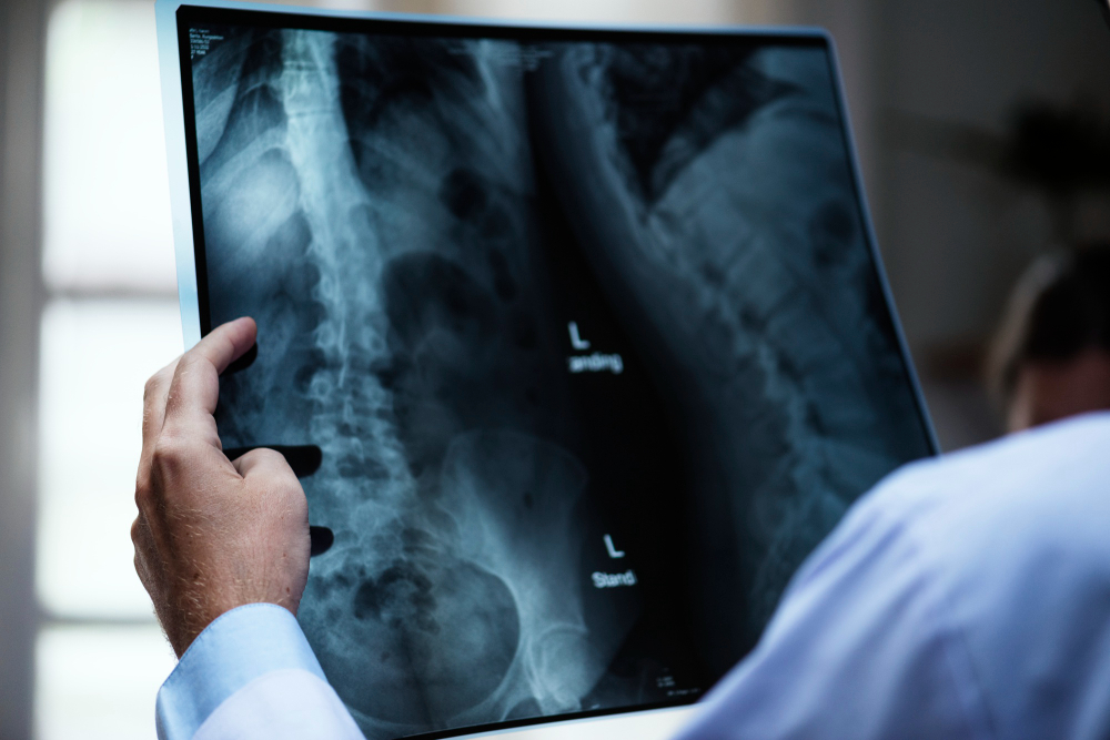
There Is No Lens, and There Cannot Be
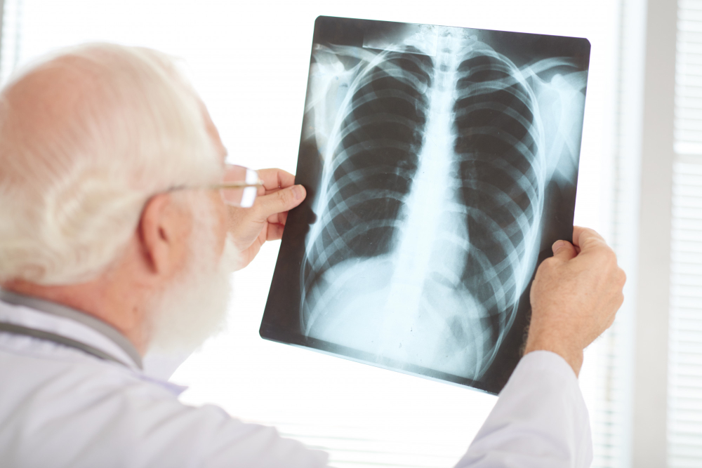
Every camera works by bending light. A lens collects rays arriving from a point on an object and bends them back together so they meet at a point on the detector, which is what makes an image rather than a smear.
X-rays cannot be bent usefully in that way. Their wavelength is far shorter than visible light and they interact with matter differently: they pass through most materials almost unaffected, and the small amount of bending that does occur is nothing like enough to focus a beam at ordinary scales. Glass is transparent to them. A lens made of anything would simply be something else they go through.
Which leaves one way to form an image, and it is the oldest way there is. Put the source on one side, the object in the middle and the detector on the other side, and record the shadow.
That is the entire optical principle of a radiograph, and almost every peculiarity of the technique follows from it. It is a contact print of a silhouette, made with a form of light that nothing can steer.
Like our content? Follow us for more.
Which Is Why the Bones Are White
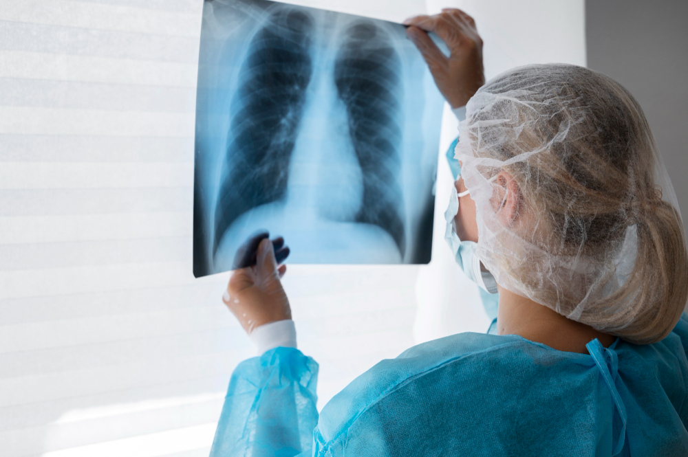
The image is a map of how much got through, and the convention makes it look inverted compared with what people expect.
Where a lot of radiation reaches the detector, the detector is strongly exposed. On the film-based system the image came from, strong exposure meant dark, so air and soft tissue came out black or grey. Where very little got through, the detector was barely exposed and the area came out pale.
So bone is white because bone is the thing that stopped the radiation, not because it emitted anything. A radiograph is a negative, and the brightest part of the picture is the part that received least.
What determines absorption is not how hard a material feels but what it is made of, specifically how heavy its atoms are. Heavier elements absorb far more strongly, and bone contains a great deal of calcium while soft tissue is mostly carbon, oxygen and hydrogen. That difference in atomic weight, not the difference in density or hardness, is why the skeleton appears so clearly against the rest of the body.
It also explains why the technique is so good at some things and so poor at others. A bone against muscle is an easy problem. Two soft tissues against each other are nearly indistinguishable, because they are made of essentially the same light elements, which is why a plain radiograph is excellent for a fracture and close to useless for most of the organs around it.
The Shadow Is Blurred for Two Reasons
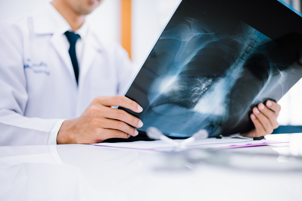
A perfect shadow needs a perfect point source. Any real source has a size, and a source with a size produces a soft edge, because different parts of it cast the shadow in slightly different places.
So one of the central design problems is making the region that emits the radiation as small as possible. That is in direct conflict with the other requirement, which is producing enough radiation, because the emitting spot gets extremely hot and a smaller spot concentrates the heat further. The entire design of the tube is a compromise between a small spot for sharpness and a large spot for survival, which is why such tubes have rotating targets: the hot area is spread around a moving ring rather than sitting in one place.
The second blur is magnification. A shadow cast by a point source is always larger than the object, and how much larger depends on the distances involved. Anything further from the detector is magnified more, so a structure at the front of the body and an identical one at the back do not appear the same size.
Which is why positioning is so exacting, and why the distances are standardised. The geometry of the shadow is part of the measurement, and changing where somebody stands changes the apparent size and shape of everything inside them.
Scatter Is the Enemy, and the Fix Is a Comb

Not everything that passes through goes straight. A proportion of the radiation is deflected sideways, carries on at a new angle, and arrives somewhere on the detector it has no business being.
That scattered radiation carries no useful information about where it came from. It arrives as a general fog across the whole image, reducing the contrast between the parts that matter, and the thicker the part of the body being examined the more of it there is.
The standard solution is a grid: a flat array of very thin strips of a heavily absorbing metal, standing on edge, separated by material the rays pass through easily, placed between the patient and the detector. Radiation travelling straight from the source passes down the channels between the strips. Radiation arriving at an angle hits the side of a strip and is absorbed.
It is a mechanical filter for direction, and it works extremely well. The cost is that it also absorbs some of the useful radiation, so using one requires a higher exposure, which is one of the standing trade-offs of the whole field: a clearer picture and more radiation, or a foggier picture and less.
Two Views, Because a Shadow Has No Depth

A shadow collapses three dimensions into two and throws away the third completely. Everything along the path of the beam is superimposed into a single point on the image, in no particular order.
So a radiograph cannot tell you how far into the body anything is. A mark could be at the front, at the back or in the middle, and it could be in front of or behind a structure it appears to overlap. A plain image simply does not contain that information.
Which is why views are taken in pairs at right angles to each other, as a matter of routine. Two shadows from two directions allow a position to be worked out by triangulation, in exactly the way two bearings fix a point on a map. A single view is frequently uninterpretable for anything where depth matters.
It is also why things can hide. A structure positioned directly behind a dense one is in its shadow and may be invisible, and a small change in angle can bring it into view. That is not a failure of the equipment; it is an unavoidable property of casting a shadow.
Contrast Media Are Borrowed Heavy Atoms
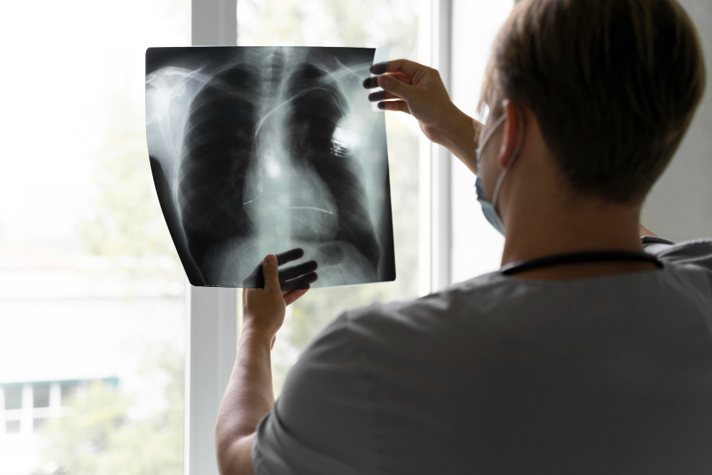
Since absorption depends on atomic weight, the way to make something visible that is not naturally visible is to fill it with heavy atoms.
That is what contrast media are. A substance containing a heavy element is introduced into a space, the space then absorbs strongly, and a structure that was previously indistinguishable from its surroundings appears as a clear silhouette. Hollow organs, blood vessels and joint spaces are all examined this way, and what is being imaged is the shape of the fluid rather than the tissue around it.
The principle is the same as throwing flour over something invisible. Nothing about the organ has changed; it has simply been coated in atoms the beam cannot get past.
This is also why the technique is entirely unlike the others that came after it. Later methods produce an image from a property of the tissue itself. A radiograph with contrast produces an image of a shape that something has been poured into, which is a different kind of picture altogether.
A Scan Is Thousands of Shadows and a Great Deal of Arithmetic

The limitation that a shadow has no depth was eventually solved, not by building a lens, but by taking an enormous number of shadows and reconstructing the object mathematically.
Rotate the source and detector around the body, record a shadow from every angle, and the question becomes: what arrangement of absorbing material would produce exactly this set of shadows? That question has a solution, it can be computed, and the answer is a cross-sectional map of how absorbing every point inside the body is.
Stack enough of those sections and you have a three-dimensional model, which can be sliced in any plane or viewed from any direction afterwards. Nothing was focused and nothing was imaged optically. The depth information was recovered by arithmetic from a collection of flat silhouettes.
That is a remarkable idea and it is worth separating from the hardware. The hardware is the same hardware: a source that cannot be focused and a detector recording shadows. What changed was realising that enough shadows contain the third dimension implicitly, and that a computer could extract it.
The cost, inevitably, is that taking many shadows means delivering more radiation than taking one, which is the central consideration in deciding between the two.
It Was a Public Spectacle Before Anybody Understood It
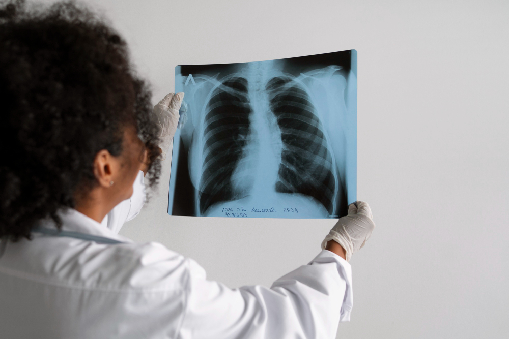
For a period after the effect was discovered, the ability to see inside a living hand was a popular entertainment. Machines appeared at fairs, in shops and in department stores, and people queued to look at their own skeletons.
Shoe shops in particular fitted devices that let a customer look at the bones of their feet inside a shoe, and these remained in use for decades after the risks were understood in principle.
The hazards were not immediately obvious because the injuries are delayed. Radiation does its damage by breaking molecules, including the ones carrying genetic information, and the consequences of that appear months or years later rather than at the time. An effect with no immediate sensation and a long delay is exactly the kind of hazard that a public with no reason to suspect it will walk straight into.
Some of the early workers in the field were injured severely, and the recognition of what was happening came from them. The subsequent history of the technique is largely a history of using progressively less of it to get progressively more information, which is why modern equipment delivers a small fraction of what early machines did for a comparable image.
None of which is advice, and nothing here is a statement about any present-day procedure. It is included because the history explains the field’s unusual preoccupation with dose, which can look excessive until you know where it came from.
A Silhouette, Then and Now
So an X-ray image is a shadow, cast by a radiation that cannot be focused by any lens anybody has managed to build, recorded as a negative in which the brightest areas are the ones that received the least.
It shows bone clearly because calcium has heavy atoms and soft tissue does not. It is blurred by the size of the emitting spot and magnified by the geometry. It is fogged by scattered radiation, which is removed by a comb of metal strips at the cost of needing more exposure. It cannot tell depth, which is why two views are taken. It can be made to show hollow spaces by filling them with heavy atoms. And it can be made to show depth after all, by taking thousands of shadows and computing what must have cast them.
Every one of those follows from the single fact at the start. There is no lens, so there is only a shadow, and everything since has been a matter of getting as much as possible out of one.
Like our content? Follow us for more.
